Welcome to the Amira-Avizo Software Use Case Gallery
Below you will find a collection of use cases of our 3D data visualization and analysis software. These use cases include scientific publications, articles, papers, posters, presentations or even videos that show how Amira-Avizo Software is used to address various scientific and industrial research topics.
Use the Domain selector to filter by main application area, and use the Search box to enter keywords related to specific topics you are interested in.
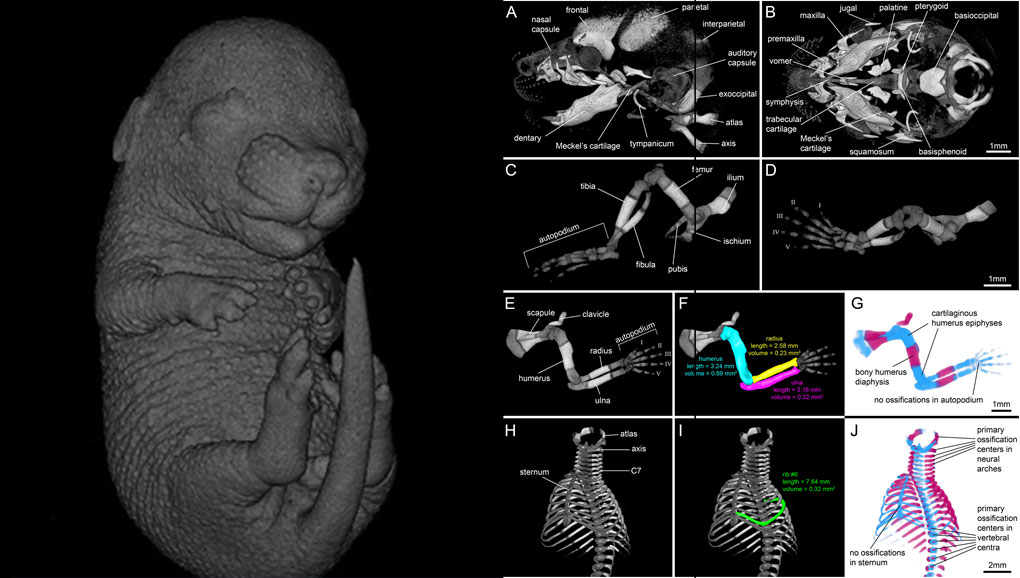
For decades, clearing and staining with Alcian Blue and Alizarin Red has been the gold standard to image vertebrate skeletal development. Here, we present an alternate approach to visualise bone and cartilage based on X-ray microCT imaging, which allows the collection of genuine 3D data of the entire developing skeleton at micron resolution.
Our novel protocol is based on ethanol fixation and staining with Ruthenium Red, and efficiently contrasts cartilage matrix, as demonstrated in wh... Read more
Simone Gabner, Peter Böck, Dieter Fink, Martin Glösmann, Stephan Handschuh
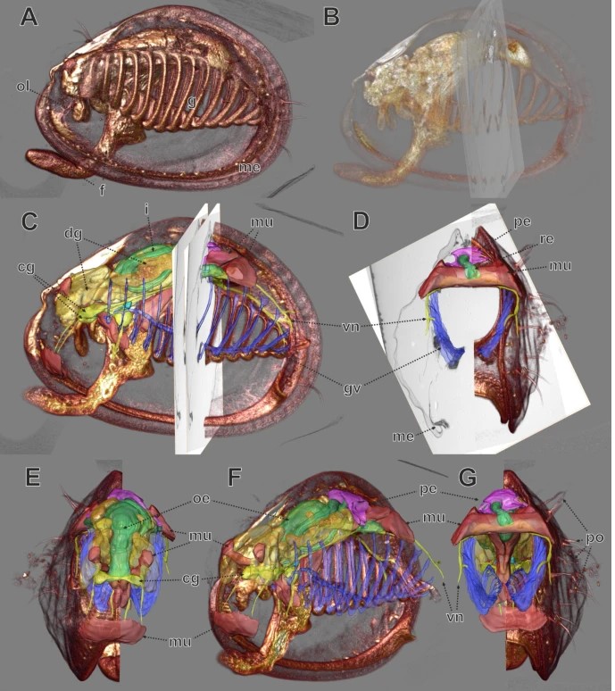
In biomedical research, a huge variety of different techniques is currently available for the structural examination of small specimens, including conventional light microscopy (LM), transmission electron microscopy (TEM), confocal laser scanning microscopy (CLSM), microscopic X-ray computed tomography (microCT), and many others. Since every imaging method is physically limited by certain parameters, a correlative use of complementary methods often yields a significant broader range of inform... Read more
Stephan Handschuh, Natalie Baeumler, Thomas Schwaha & Bernhard Ruthensteiner
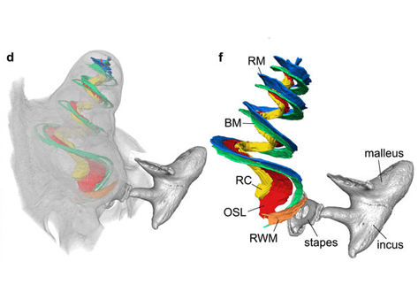
Propagation-based phase-contrast x-ray tomography of cochlea using a compact synchrotron source
We demonstrate that phase retrieval and tomographic imaging at the organ level of small animals can be advantageously carried out using the monochromatic radiation emitted by a compact x-ray light source, without further optical elements apart from source and detector. This approach allows to carry out microtomography experiments which – due to the large performance gap with respect to conventional laboratory instruments – so far were usually limited to synchrotron sources. We dem... Read more
Mareike Töpperwien, Regine Gradl, Daniel Keppeler, Malte Vassholz, Alexander Meyer, Roland Hessler, Klaus Achterhold, Bernhard Gleich, Martin Dierolf, Franz Pfeiffer, Tobias Moser & Tim Salditt
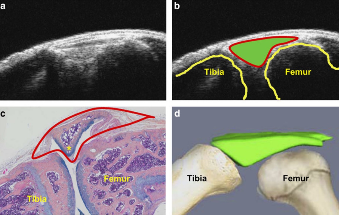
Ultrasound could be a fast and cost-effective means of assessing joint changes in mouse models of posttraumatic osteoarthritis (PTOA). Such models are essential for understanding the biology of this degenerative joint disease and developing new treatments, but noninvasive methods of evaluating disease activity are lacking. Because ultrasound can visualize both joint space volumes and blood flow in the joints, it could provide an alternative to microscopic examination of tissue, assuming it ac... Read more
Hao Xu, Echoe M Bouta, Ronald W Wood, Edward M Schwarz, Yongjun Wang & Lianping Xing